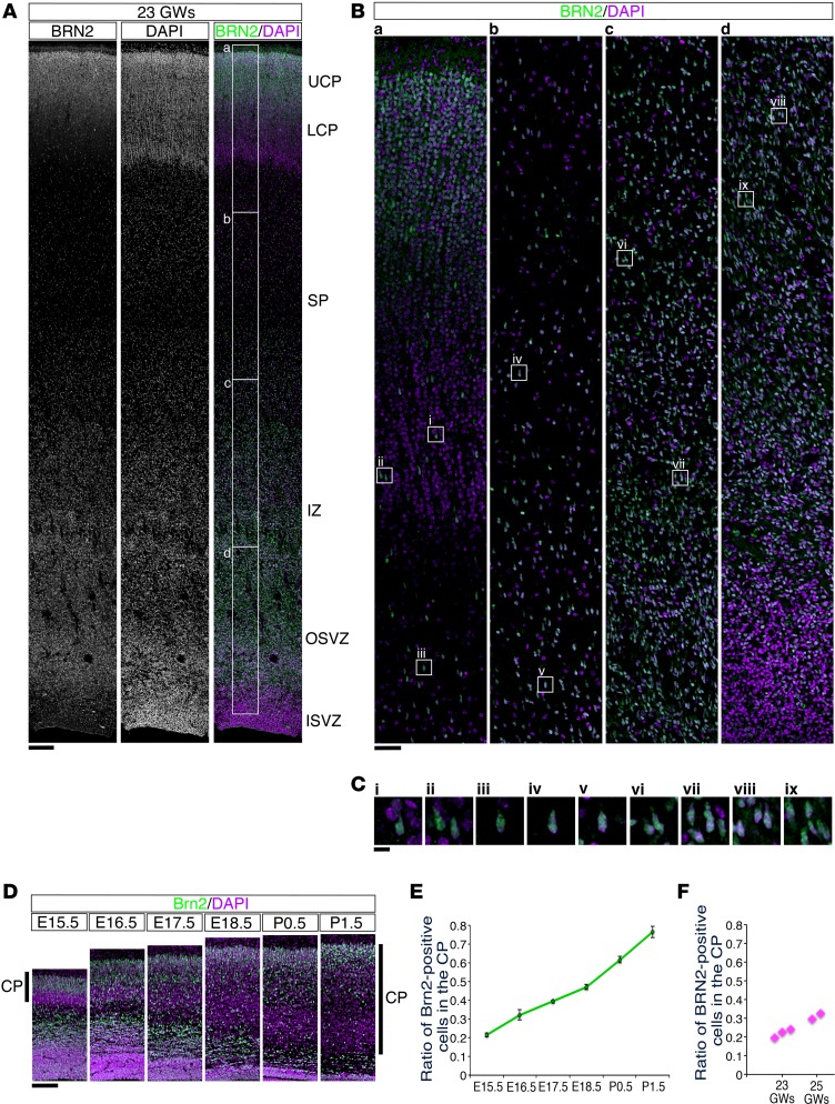
Figure 4. BRN2-positive cells were abundantly distributed in the subplate and intermediate zone of the human neocortex at 23 GWs.
(A) Sections from the human occipital neocortex at 23 GWs were stained with anti-BRN2 antibody (left; gray scale, right; green) and counterstained with DAPI (middle; gray scale, right; purple). (B) High-magnification images of the boxed areas in A (labeled a–d) are shown. The BRN2-positive cells accumulated at the top of the cortical plate (CP) and were diffusely distributed throughout the neocortex, including the subplate (SP) and intermediate zone (IZ). (C) High-magnification images of the squares in B (labeled i–ix) are shown. Scale bar: 200 μm (A); 50 μm (B); 10 μm (C). (D) Sections from the developing mouse neocortex from E15.5 to P1.5 were stained with an anti-Brn2 (green) antibody and counterstained with DAPI (purple). Scale bar: 200 μm. (E) The ratio of the number of Brn2-positive cells in the CP to the total number of Brn2-positive cells throughout the mouse neocortical wall was calculated (mean ± SEM; n = 4, respectively). (F) The ratio of the number of BRN2-positive cells in the CP to the total number of BRN2-positive cells throughout the human neocortical wall (23 GWs: n = 3, 25 GWs: n = 2) was calculated and plotted.

