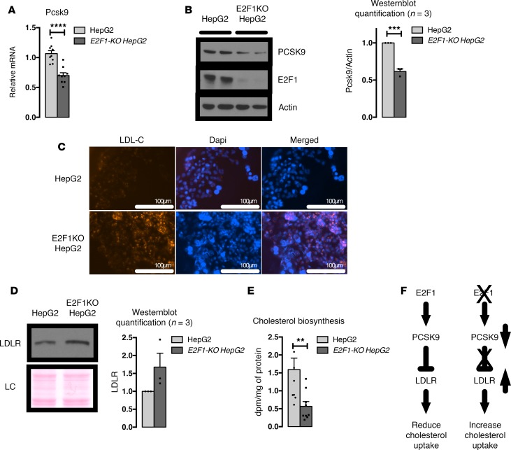
Figure 4. E2F1-KO HepG2 cells show increased LDL-cholesterol (LDL-C) uptake and decreased cholesterol biosynthesis.
(A) Relative expression of PCSK9 mRNA in HepG2 and E2F1-KO HepG2 cells. Three independent experiments in triplicate. (B) Representative images of PCSK9 Western blots from HepG2 and E2F1-KO HepG2 cells. Three independent experiments. Normalization of the quantified values is represented. (C) Representative images of LDL-C uptake in HepG2 and E2F1-KO HepG2 cells. Three independent experiments. Red fluorescence denotes LDL-C, and nuclei are stained in blue. Scale bars: 100 μm. (D) Representative images of LDLR Western blots from HepG2 and E2F1-KO HepG2 cell membranes. Three independent experiments. Ponceau staining is shown as loading control (LC). (E) Quantification of the rate of cholesterol biosynthesis in HepG2 and E2F1-KO HepG2 cells. Three independent experiments in triplicate. (F) Representative role of E2F1 in PCSK9-LDLR cholesterol uptake. All data are presented as the mean ± SEM. Differences between HepG2 and E2F1-KO HepG2 were determined by 2-tailed unpaired t test. **P < 0.01, ***P < 0.001, ****P < 0.0001.

