
Fig. 1.
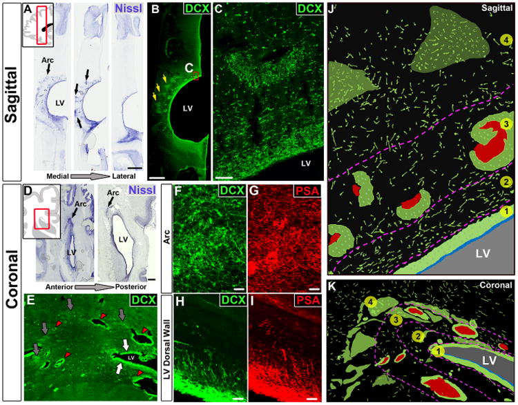
Migrating young neurons in the infant frontal lobe are widely distributed in four tiers. (A) Serial Nissl-stained sections (taken at birth) reveal cell-dense collections around the anterior body of the lateral ventricle (black arrows, defined here as the Arc); LV, lateral ventricle. (B and C) The cells in these densities (yellow arrows) and next to the ventricular wall express DCX. (D) Coronal sections (38 GW) showing cell densities close to the ventricular wall (eyebrow-shaped, black arrows). (E) Dense aggregates of DCX+ cells around the walls of the lateral ventricles (white arrows), around blood vessels (red arrowhead), and in the parenchyma within the Arc (gray arrows). (F to I) DCX+ cells also express PSA-NCAM; (F) and (G) show cells within the Arc; (H) and (I) show cells next to the ventricular walls. (J and K) Schematic drawings of traced DCX+ cells (in green) illustrating how cells within the Arc are organized into four tiers (see text). Blood vessels are shown in red; light green clusters correspond to DCX+ cellular densities seen in (B) and (E). Scale bars, 2 mm [(A) and (B)], 50 μm (C), 1 mm (D), 25 μm [(F) to (I)].