
Fig. 6. Migratory streams of young neurons in the frontal lobe of the early postnatal human brain.
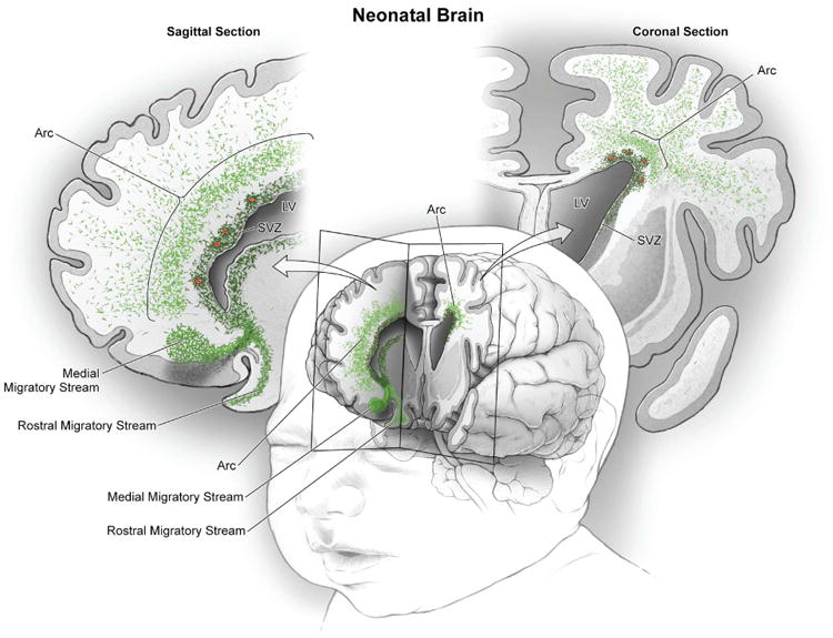
In the frontal lobe of the neonatal human brain, cut in sagittal and coronal planes in this schematic, large numbers of young migrating neurons persist (shown in green) (see Figs. 1 to 3). Multiple concentric tiers of migrating cells are observed around the anterior pole of the lateral ventricle (see Fig. 1). Close to the ventricular wall, migrating young neurons are largely oriented tangentially; dense subpopulations are also clustered around blood vessels (red). Farther out, young neurons are more dispersed, many now oriented radially; they appear to migrate long distances through the developing white matter to reach the cortex. Ventrally, we also illustrate the RMS and the MMS, which target the olfactory bulb and medial prefrontal cortex, respectively (20).