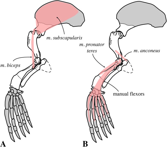
Figure 29. Fore limb musculature of Nanophoca vitulinoides in medial view.
The origin and insertion of selected muscles of the fore limb that are visible in medial view. Muscles indicated in pink. Missing bones or bone parts of Nanophoca vitulinoides indicated in gray. Dashed line visually completes the ulna. This illustration focuses on the visualization of the origin and insertion of different muscles. Hence, the actual shape of the muscles may differ from this illustration.

