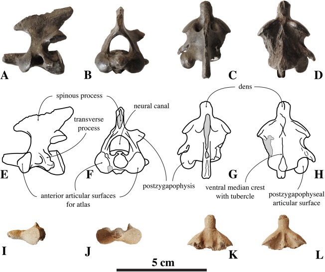
Figure 3. Axes of Nanophoca vitulinoides.
IRSNB M2268 axis of Nanophoca vitulinoides (A–D) and corresponding drawings (E–H) in left lateral (A, E), anterior (B, F), dorsal (C, G), and ventral (D, H) view. IRSNB M2276i (neotype) axis of Nanophoca vitulinoides in left lateral (I), anterior (J), dorsal (K), and ventral (L) view. Broken or obliterated areas are indicated in gray.

