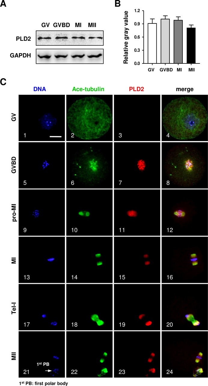
Figure 1. The protein expression of PLD2 and its relationship with spindle in mouse oocytes during meiotic maturation.
(A) Western blot analysis detected stable expression of PLD2 at the GV, GVBD, MI and MII stages, respectively. The experiments were repeated triply. (B) Statistical analysis confirmed no significant difference in PLD2 expression among germinal vesicle (GV), germinal vesicle breakdown (GVBD), metaphase I (MI) and metaphase II (MII) stages (P > 0.05). (C) PLD2 was co-localized with spindle during meiotic division. At the GV stage, no particular aggregation of PLD2 was detected throughout the cytoplasm and nuclear (1–3). After GVBD, PLD2 emerged as filamentous assembly and was co-localized with newly formed microtubules around the condensed chromosomes (5–8). From pro-MI to MI, PLD2 was co-localized with the spindle structure (9–16), and during anaphase I (AI) to telophase I (Tel I), it was mainly distributed at spindle poles and absent from the midbody area (17–20). At the MII stage, PLD2 was again co-localized with microtubules on the re-formed meiotic spindle and first polar body (1st PB) (21–24: arrow). DNA was visualized in blue, microtubules were in green and PLD2 was in red. Scale bar = 20 µM.

