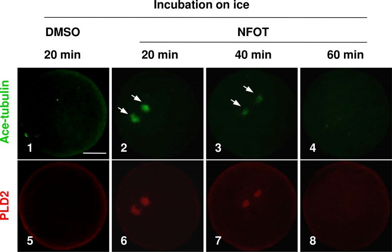
Figure 3. The spindle microtubules were stabilized in oocytes with PLD2 inhibition with NFOT.
GV oocytes were incubated for 8 h with DMSO or NFOT, and then collected for additional incubation in fresh M2 medium on ice for 20 min, 40 min and 60 min, respectively, and followed by immunofluorescence procedure. The experiment was repeated triply with more than 30 oocytes analyzed in each group every time. Microtubules were labeled in green and PLD2 in red. Scale bar = 20 µM. At 20 min of cooling treatment, the microtubules were totally disassembled together with PLD2 in control oocytes; however, both microtubules and PLD2 remained in NFOT-treated oocytes at this time, even sustained at 40 min, and completely vanished at 60 min.

