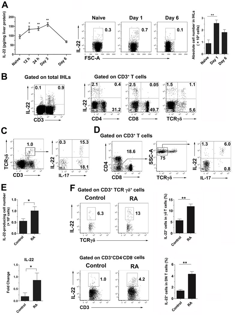
Figure 2. RA promoted IL-22 expression by γδ T cells and double negative (DN) T cells in the liver of viral hepatitis.

B6 mice were i.v. injected with 3 × 10⁹ pfu of AdLacZ and sacrificed at the indicated time points. (A) Kinetic analysis of hepatic IL-22 production by an ELISA assay. IHLs were stimulated with recombinant IL-23 (rIL-23, 20 ng/ml) for 16 h plus GolgiStop for the last 4 h. The cells were examined for intracellular IL-22. Right panel: Cumulative statistical results of the absolute number of IL-22+ cells in the liver. (B) IHLs from day 1 infected mice were gated on the CD3+ population. IL-22-producing cells were further analyzed on CD4+, CD8+ and TCRγδ+ T cells. (C) The IHLs were gated on γδ T cells for IL-17 and IL-22 expression. (D) The IHLs were gated on DN T cells (CD3+ CD4− CD8− TCRγδ− cells) for IL-17 and IL-22 expression. (E) Mice were infected and treated with RA as shown in Fig. 1. Hepatic IL-22-producing cells were analyzed at 6 dpi. Left panel: absolute number of IL-22+ cells. Right panel: IL-22 mRNA level of IHLs. (F) Intracellular IL-22 expression in hepatic γδ T and DN T cells. The experiment was repeated two to three times independently, and a representative graph is shown (6–8 mice/group). *p < 0.05, **p < 0.01.
