
Figure 3. IL-17 and IL-22 production in γδ T and DN T cells was dependent on the PI3K/mTORC1 signaling pathway.
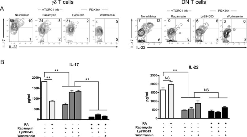
B6 mice were i.v. injected with 3 × 10⁹ pfu of AdLacZ and sacrificed at 2 dpi. IHLs were isolated and stimulated with rIL-23 (20 ng/ml) for flow cytometry. The culture system was initially added with or without RA (100 nM), Rapamycin (25 nM), Ly294002 (5 μM) or Wortmannin (100 nM). DMSO was used as solvent control. For intracellular staining, the cells were cultured for 16 h plus GolgiStop for the last 4 h. (A) γδ T and DN T cells were gated first, followed by the analysis of intracellular IL-17 and IL-22 expression. (B) For the detection of cytokines in the supernatant, the cells were cultured for 3 days. The supernatants were collected for an ELISA assay. The experiment was repeated three times independently. **p < 0.01, ***p < 0.001.