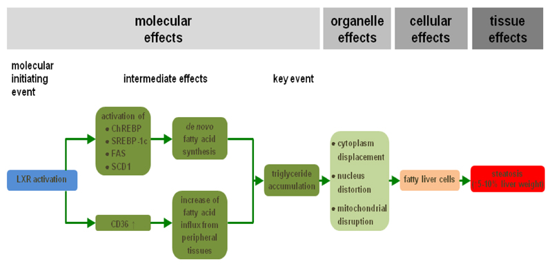
Figure 2. AOP for drug-induced liver steatosis.
Activation of the liver X receptor (LXR), which is the MIE (blue), induces a number of transcriptional changes, including activation of the expression of carbohydrate response element binding protein (ChREBP), sterol response element binding protein 1c (SREBP-1c), fatty acid synthase (FAS) and stearoyl-coenzyme A desaturase 1 (SCD1). As a result, de novo synthesis of fatty acids is enhanced in the liver. At the same time, fatty acid translocase (CD36) production is upregulated, which mediates increased hepatic influx of fatty acids from peripheral tissues. All together, these intermediate steps drive accumulation of triglycerides, which is considered a key event (dark green). At the organelle level, this evokes cytoplasm displacement, distortion of the nucleus and mitochondrial disruption. This ultimately burgeons into the appearance of fatty liver cells (orange) and further into the clinical diagnosis of liver steatosis (red) (adapted from (20)).

