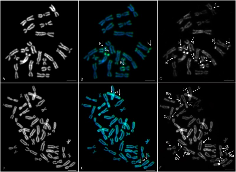
Fig 1. Sequential fluorescent chromosome banding on metaphase spread of X. tropicalis and X. mellotropicalis.
DAPI (B&W) counter-stained metaphase spreads showed all (A) 20 X. tropicalis and/or (D) 40 X. mellotropicalis chromosomes. (B) CMA3 (green) and (C) C-banding (B&W) in X. tropicalis stained the part of q arms of XTR 9 and p arms of XTR 3. Moreover, (C) the pericentric region of XTR 4 and 10 and p arms of XTR 8 were weakly stained by C-banding. X. mellotropicalis chromosomes stained by (E) CMA3 (green) and (F) C-banding (B&W) revealed positive bands located on the p arm pericentric region of XME 2a. In addition, (F) C-banding detected further minor heterochromatic blocks on X. mellotropicalis chromosomes 1b, 2b, 6b, 7b, 8a and 10a. (B, C, E, F) All arrows show heterochromatic blocks. Scale bar represents 10 μm.

