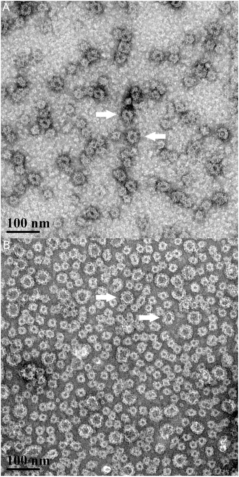
Fig 1. Transmission electron microscopy of norovirus VLPs.

GI (A) and GII.4 (B) VLPs were dissolved in water and imaged at 150,000x magnification (scale bar 100 μm). VLP particles were spherical in appearance at the expected size of 23–38 nm.

GI (A) and GII.4 (B) VLPs were dissolved in water and imaged at 150,000x magnification (scale bar 100 μm). VLP particles were spherical in appearance at the expected size of 23–38 nm.