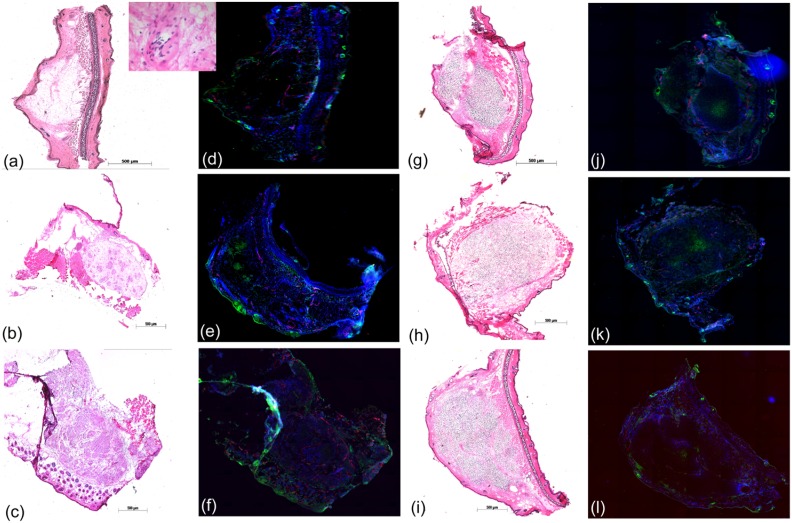
Fig 5. Histology of the small mouse ear tumours.
Exemplary tumour sections used to characterize the tumour micromileu for tumour sizes of 1–2 mm (1st line), ≤ 2.5 mm (2nd line) and at treatment size of 3–4 mm (3rd line) for HNSCC FaDu and GBM LN229. The tumour appearance within the surrounding normal tissue was confirmed by classical H&E staining (a-c, g-i) and is shown in 50x magnification, except the inset of Fig 5a, where a magnification of 400x was applied. The analysis of tumour histology takes place by immunofluorescence staining of perfused areas (blue, Hoechst 33342), of small vessels (red, CD31) and of hypoxic regions (green, pimonidazole). Due to technical reasons sections of two different tumours are shown in Fig 5b and 5e, whereas the stained sections of all other pairs were taken from the same tumour.

