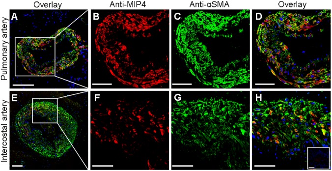
Fig 3. Colocalization of MIP-4/PARC and alpha smooth muscle actin (αSMA) in arterial tissue.
Representative confocal fluorescence images of human pulmonary muscular (A) and intercostal (E) arteries labeled with antibodies against MIP-4/PARC (red) and αSMA (green). Nuclei were stained in blue. Higher-magnification (x40 oil lents) of representative images shows that MIP-4/PARC (B and F) and αSMA (C and G) were predominantly localized in the media layer of pulmonary and intercostal arteries. Overlay images show, in yellow, a partially colocalization between MIP-4/PARC and αSMA (D and H). Inset in H corresponds to control experiments performed with secondary antibodies alone. Scale bars = 50 μm except for images A and E, where they are 100 μm.

