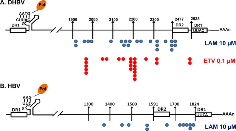
Figure 8. Mapping the 3′ ends of encapsidated viral RNA.

(A) dstet5 cells were treated with 10 μM LAM or 0.1 μM ETV for 3 days in the absence of tet. (B) HepAD38 cells were treated with 10 μM LAM for 5 days in the absence of tet. Encapsidated polyA-free viral RNAs were extracted and their 3′ termini were determined by a 3′-RACE procedure as described in Materials and Methods. The position of 3′ terminus of each of the sequenced DHBV or HBV cDNA was depicted in the diagram of viral pgRNA.
