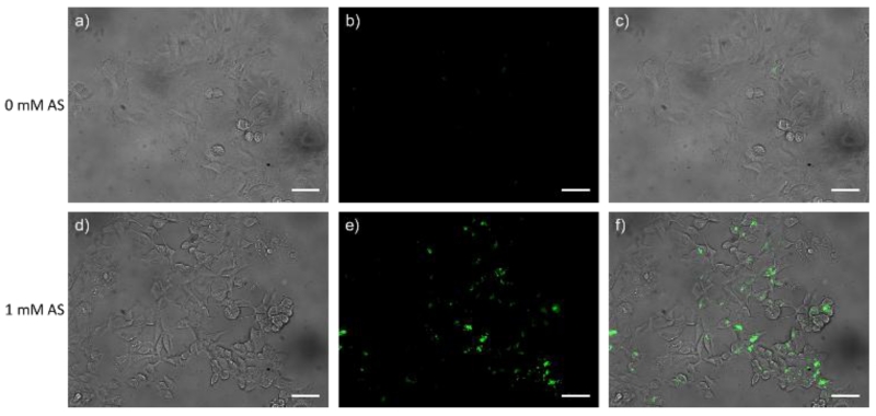
Fig. 5.
Fluorescence images of live MCF-7 cells. (a) bright-field image of living MCF-7 cells incubated with only Pdot-PFBT/PC30-Cu2+; (b) fluorescence image of (a); (c) merged image of (a) and (b). (d) bright-field image of living MCF-7 cells incubated with Pdot-PFBT/PC30-Cu2+ and 1 mM AS; (e) fluorescence image of (d); (f) merged image of (d) and (e). The scale bar is 50 μm.

