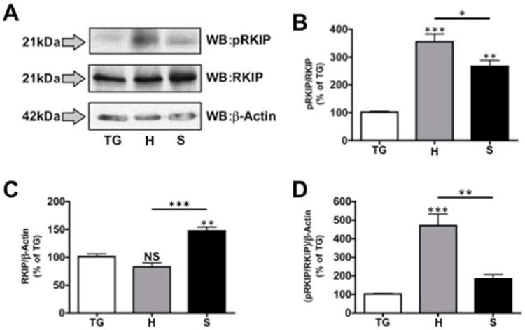
Figure 1. RKIP phosphorylation and expression in oral cancer cell lines.

Primary trigeminal neuron cultures (TG), HSC3 (H) and SCC4 (S) cell lines were lysed and prepared for proteomic analysis by Western blot. A. Proteins were identified using antibodies specific for phospho Ser153 RKIP (pRKIP), total RKIP (RKIP) and β-Actin. Protein molecular weights are shown to the left of the lots. Densitometry was performed on immunoreactive bands, with pRKIP normalized to total RKIP (B), total RKIP normalized to β-Actin (C) and pRKIP normalized to RKIP/β-Actin (D). Results shown are representative of 3 independent trials; statistics determined by one-way ANOVA with Bonferroni post-hoc correction.
