
Figure 5. Effect of gel stiffness on the formation of pancreatic cancer cell spheroids.
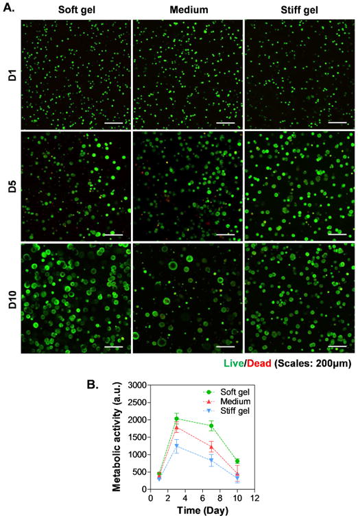
(A) Representative confocal z-stack images of live/dead stained COLO357 cells. (B) Cell metabolic activity as assessed by Alamarblue® reagent. All gel formulations contained 7 wt% GelNB, 0.9 wt% THA, as well as 0, 1 or 2 wt% of PEG4SH (i.e., soft, medium and stiff hydrogel).