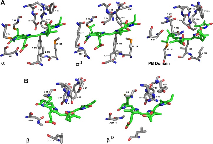
Fig 5. Binding sites of phycocynobilin.
A) Sticks representation of binding site of phycocyanobilin in α subunit of 5TJF (this paper), αII and the PB domain in APC_3. The chromophores are represented in green. B) Sticks representation of β subunit in 5TJF and β18 in APC_3. The chromophores are also in green.

