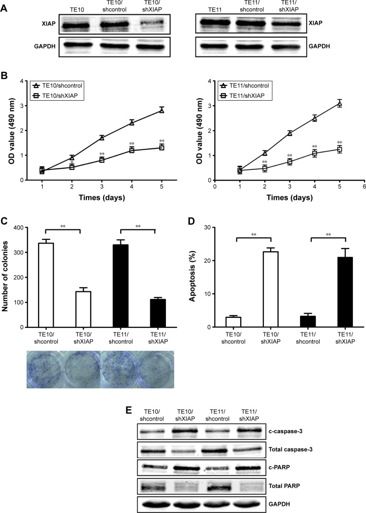
Figure 5.
Effects of XIAP knockdown on growth, colony formation, and apoptosis in ESCC cells.
Notes: (A) Western blot of XIAP protein expression in TE10 and TE11 cells stably transfected with pGLV3/shXIAP and pGLV3/shcontrol, respectively. GAPDH was used as an internal control. (B) MTT analysis of growth in TE10 and TE11 cells stably transfected with pGLV3/shXIAP and pGLV3/shcontrol, respectively. (C) Colony formation assay was performed. (D) Flow cytometric detection of apoptosis in TE10 and TE11 cells stably transfected with pGLV3/shXIAP and pGLV3/shcontrol, respectively. (E) Western blot detection of c-caspase-3, total caspase-3, c-PARP, and total PARP proteins in the stably transfected TE10 and TE1. GAPDH was used as an internal control. Each experiment was performed at least in triplicate. *P<0.05 and **P<0.01 vs control.
Abbreviations: XIAP, X-linked inhibitor of apoptosis protein; ESCC, esophageal squamous cell carcinoma; OD, optical density.

