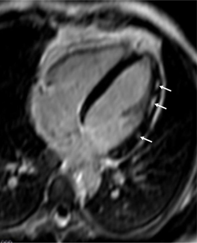
Figure 4.

Patchy fibrosis due to myocardial inflammation in a patient with polymyositis.
Note: The arrows show areas of patchy fibrosis in the lateral wall of LV, due to myocardial inflammation in a patient with polymyositis.
Abbreviation: LV, left ventricle.
