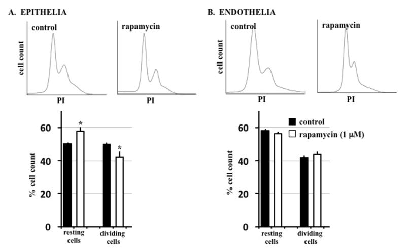
Figure 6. Treatment with rapamycin shows a minimal effect on the cell growth.

Cells were stained with propidium iodide (PI), a DNA intercalating agent, for flow cytometry analysis. (A) Epithelial cells showed a slight but significant increase in cells at the resting (G0) stage and lower percentage of cells undergoing mitosis when treated with rapamycin. (B) For endothelial cells, no significant variation in cell cycle was seen in rapamycin-treated cells compared to control. *= p<0.05 compared to corresponding controls.
