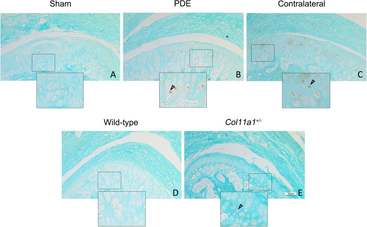
Fig 2. Increase in the protein expression of p-Smad2/3 in condylar cartilages in mice.
There were p-Smad2/3 protein positive staining cells (brown-color staining) in the condylar cartilage of discectomy mice (B), Col11a1+/- mice (E) and the contralateral non-surgical TMJ of mice (C). The expression patterns of p-Smad2/3 were similar to what were seen in the elevated expression of Tgf-β1 in the Fig 1 above. The positive-staining cells were seen within the basal layer of the condylar cartilages. There were no positive staining cells seen in sham control (A) and wild-type mice as control (D). (Bar = 50 μm).

