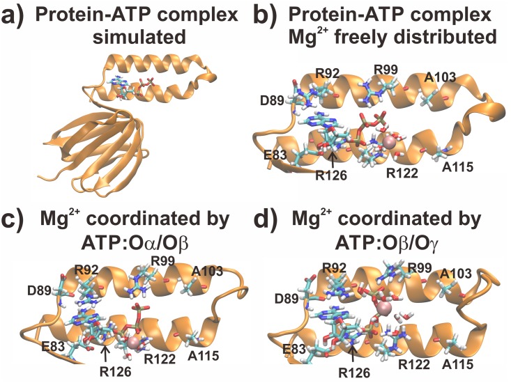
Fig 1. Input structures for the simulations.
In a) the whole protein-ATP complex is shown. In b-d) the initial binding site structures for the freely distributed Mg2+ case, the MgATP:Oα/Oβ and MgATP:Oβ/Oγ coordination are shown, respectively. Water molecules coordinating the Mg2+ ion and residues coordinating ATP are shown in licorice, while the Mg2+ ion is shown in VdW spheres.

