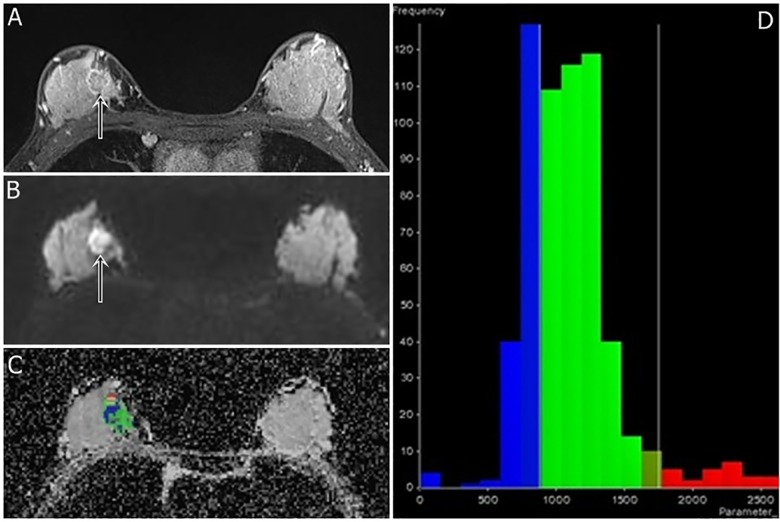
Fig 2. A 41-year-old woman with triple-negative cancer in the right breast (poorly differentiated grade, high Ki-67 index, and high ADC kurtosis).
Fat-suppressed contrast-enhanced T1-weighted imaging (a) shows an irregular rim-enhancing mass (arrow). A DWI (b = 750 s/mm2) (b) shows positive rim sign (arrow). DWI slice of tumor volume reconstruction of ADC values (c) and a histogram map (d) are shown. ADC mean; mode; and 25th, 50th, and 75th percentiles were 1.099, 0.745, 0.859, 1.075, and 1.245 x 10⁻³ mm2/s, respectively. The ADC skewness and kurtosis were 1.59 and 4.28, respectively. The 3-cm-sized tumor had a poor histologic grade and high Ki-67 index (50%).

