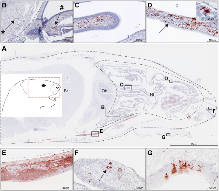
Fig 5. rCDV was detected in the nasal cavity of directly inoculated ferret D4.
Immunohistochemistry detecting CDV was performed on (A) complete ferret head sections (red box in inset shows anatomic location of section) and showed abundant rCDV in the nasal turbinates (Nt, delineated by dotted line). In this ferret rCDV was only observed in few meningeal cells and not in the cerebrum (Br) and olfactory bulb (Ob). In general, CDV-positive cells were mainly found in the submucosa of the respiratory and olfactory mucosa and nasal-associated lymphoid tissues (NALT). (B) Cells surrounding the nerve twigs of the olfactory nerve were CDV-positive on both sides of the cribriform plate (dotted line), on both the side of the nasal cavity (#) and the olfactory bulb (*). (C) Few CDV-positive cells were detected in the olfactory epithelium, and many CDV-positive cells were present in the submucosa of the olfactory epithelium. Positive cells had an irregular macrophage-like morphology. (D) CDV-positive cells were detected in the respiratory epithelium of the nasal turbinates, although most positive cells within the epithelium appear non-epithelial (arrows and insert). Again, CDV-positive cells were abundant in the submucosa and had an irregular macrophage-like morphology. (E) Positive stretch of CDV-positive cells, including NALT with infected lymphocytes and fibroblasts. CDV-positive macrophages were also detected in the epithelial layer. (F) Submucosal nasal glands stained occasionally positive for CDV predominantly in the tip of the nose (arrow). (G) Squamous epithelium of the soft palate stained positive for rCDV.

