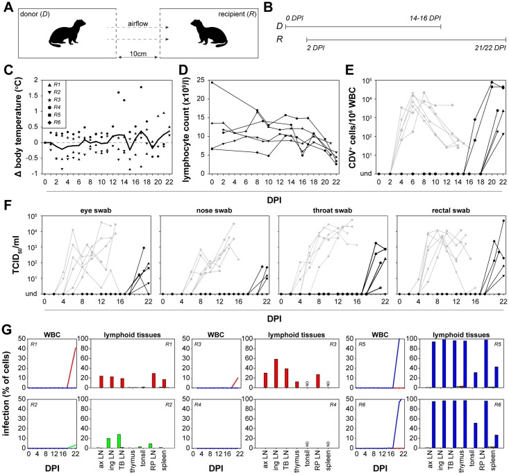
Fig 6. In vivo transmission of rCDV to recipient ferrets.
(A, B) Susceptible recipient ferrets (R1-6) were placed pairwise in neighboring cages of the donor ferret at 2 DPI. Arrows indicate airflow. All directly inoculated ferrets were euthanized at 14 to 16 DPI, whereas recipient ferrets were euthanized 21 or 22 DPI. (C) Fever was rarely detected in recipient ferrets, but these animals developed (D) lymphopenia and (E) systemic virus replication as detected by virus isolation from WBC as well as (F) local virus replication demonstrated as virus isolation from eye, nose, throat and rectal swabs. In panels E and F, grey lines represent the respective virus loads in donor ferrets (as shown in Fig 2). Transmission was detected in 6/6 animal pairs. (G) WBC infection percentages of the rCDVs in recipient ferrets were determined by flow cytometry at different time-points. After necropsy, infection percentages of the rCDVs in single cell suspensions from lymphoid tissues were determined. rCDV was detected in most lymphoid tissues (ax LN: axillary LN; ing LN: inguinal LN; TB LN: tracheo-bronchial LN; RP LN: retropharyngeal LN; ND: not determined). Results obtained from lymph nodes corresponded to the kinetics of the rCDVs in WBC. In 6/6 ferrets the single-color virus dominant in the donor animal transmitted to the recipient ferret.

