
Figure 6. α4 integrin-dependent macrophage-adipocyte interactions inhibit UCP1 expression in a phospho-Erk dependent manner.
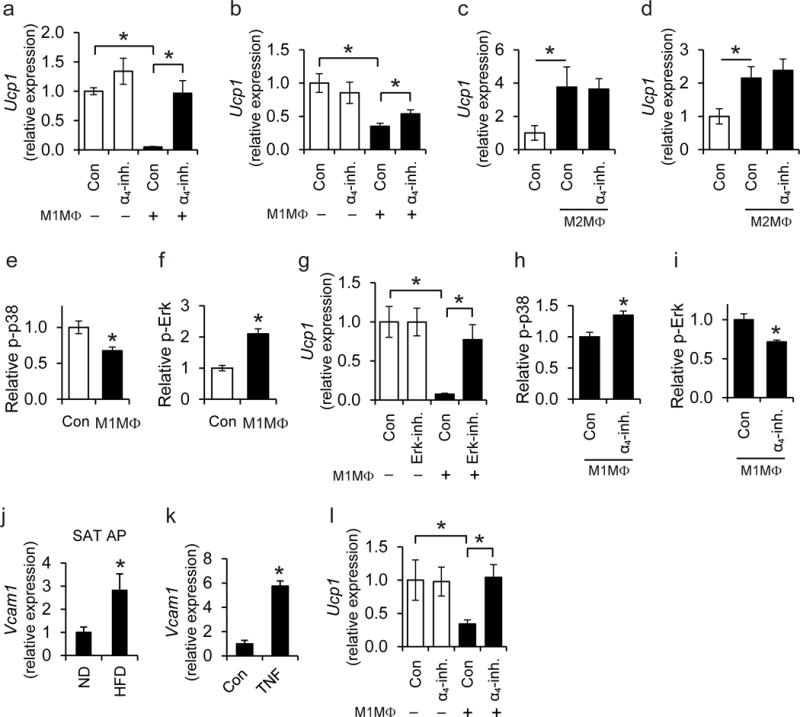
a–b) Primary adipocytes were cultured with T3 and norepinephrine to induce a thermogenic response, prior to co-culture in the absence or presence of (a) M1-like polarized pro-inflammatory BMDMs or (b) M1-like polarized primary SAT-derived macrophages [M1MΦ in the figure are M1-like polarized BMDMs in (a) or SAT macrophages in (b)] for 1 h. Co-culture was performed in direct contact (in the same well). Experiments were performed in the absence (control; Con) or presence of α4-inhibitor (α4-inh.). a) Ucp1 mRNA expression was analyzed in adipocytes after separation from the BMDMs. 18S expression was used for normalization and Ucp1 expression in adipocytes cultured in the absence of macrophages and in the absence of α4-inhibitor was set as 1 (n=5 separate primary adipocyte isolations). b) The mRNA expression of Ucp1 was analyzed in adipocytes after separation from SAT macrophages. l8S expression was used for normalization and Ucp1 expression in adipocytes cultured in the absence of macrophages and in the absence of α4-inhibitor was set as 1 (n=5 separate primary cell isolations from SAT). c–d) Primary adipocytes (pre-stimulated with T3 and norepinephrine) were co-cultured in the absence or presence of M2-like polarized primary SAT-derived macrophages (M2MΦ) in direct contact (c, in the same well) or in an indirect way (d, in transwell co-culture system, wherein adipocytes and macrophages were in the lower and upper compartment, respectively). Experiments were performed in the absence (control; Con) or presence of α4-inhibitor (α4-inh.). The mRNA expression of Ucp1 was analyzed in adipocytes after separation from the macrophages. l8S expression was used for normalization and Ucp1 expression in adipocytes cultured in the absence of macrophages was set as 1 (n=5 separate primary cell isolations from SAT). e–f) Primary adipocytes (pre-stimulated with T3 and norepinephrine) were co-cultured in the absence (control; Con) or presence of M1-like polarized BMDMs (direct contact) for 30 min. The amounts of phospho-p38 (e) and phospho-Erk (f) in adipocytes were detected by flow cytometry. Phospho-p38 or phospho-Erk protein amounts (MFI) in adipocytes cultured in the absence of M1MΦ were set as 1 (n=4 separate primary adipocyte isolations). g) Primary adipocytes were pre-treated with T3 and norepinephrine for 3 h, washed and then incubated in the absence (control; Con) or presence of an Erk inhibitor for 30 min. After washing, direct co-cultures between adipocytes and M1-like BMDMs were performed for 1 h and the mRNA expression of Ucp1 was studied in adipocytes after separation from the macrophages. The mRNA expression of Ucp1 in the absence of BMDMs and without Erk-inhibitor pre-treatment was set as 1 (n=4 separate primary adipocyte isolations). h, i) T3- and norepinephrine-pre-treated adipocytes were co-cultured with M1-like BMDMs (M1MΦ) in direct contact for 30 min in the absence (control; Con) or presence of α4-inhibitor (α4-inh.). Phospho-p38 (h) and phospho-Erk (i) in adipocytes were detected by flow cytometry and their respective amount (MFI) in the presence of M1MΦ but in the absence of α4-inhibitor was set as 1 (n=4 separate primary adipocyte isolations). j) Primary adipocyte progenitor cells (AP) were isolated from SAT of wild-type C57BL/6 lean (ND, n=4 mice) and obese (HFD, n=6 mice) mice by FACS-sorting (AP cells were defined as CD31−CD45−Sca1+PDGFRα+ cells) and Vcam1 mRNA expression was determined. 18S expression was used for normalization and Vcam1 expression of ND-fed mice was set as 1. k) AP were isolated from SVF of SAT of wild-type mice with FACS-sorting (CD31−CD45−Sca1+PDGFRα+ cells). Cells were treated without (control; Con) or with TNF. Vcam1 mRNA expression was analyzed. 18S expression was used for normalization and Vcam1 expression of control-treated cells (Con) was set as 1 (n=4 separate cell isolations). l) AP were isolated as in (k) and treated with T3 and norepinephrine. Thereafter, AP were co-cultured in the absence or presence of M1-like polarized pro-inflammatory BMDMs (M1MΦ) in direct contact with each other. Experiments were performed in the absence (control; Con) or presence of α4-inhibitor (α4-inh.). The mRNA expression of Ucp1 in AP was detected after macrophage separation. 18S expression was used for normalization and Ucp1 expression in the absence of macrophages and in the absence of α4-inh. was set as 1 (n=5 separate primary adipocyte progenitor isolations).
Data are presented as mean ± SEM. Data in (b), (c), (d) are pooled from 3 experiments; data in (a), (e), (f), (g), (h), (i), (k) are representative of 2 experiments; data in (j) and (l) are from one experiment. *P < 0.05. Mann-Whitney U-test in (a)–(l).