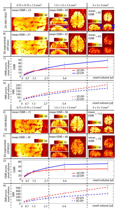
Fig. 2. tSNR across resolutions.
Results of VASO tSNR for 0.75 mm, 1.5 mm and 3 mm resolutions are shown for one participant in (A)–(B). For the protocols with (low) resolutions of 1.5 mm and 3 mm, 2D-SMS VASO provides higher tSNR values compared to 3D-segmented-EPI. At sub-millimeter resolutions of 0.75 mm, however, the tSNR of 3D-segmented-EPI surpasses that of 2D-SMS EPI. For best visibility, the dynamic range of the color bars is adjusted for each resolution, but it is kept identical for 2D-SMS and 3D-segmented-EPI. tSNR results in (C) refer to experiments, where slice-acquisition parameters are kept the same but the slice thickness is varied. Different voxel volumes refer to different slice thicknesses. It can be seen that in the physiological-noise-dominated regime of voxel volumes with few microliters, 2D-SMS-EPI performs better than 3D-segmented-EPI. However, in the thermal-noise-dominated regime at sub-microliters resolutions, 3D-segmented-EPI has higher signal stability. Fig. (E)–(H) show the same for the BOLD contrast. Similarly to VASO, also the BOLD stability is better for 3D-segmented-EPI at ultra high resolutions, while 2D-SMS is better for conventional resolutions. Note that the ceiling effect is slightly stronger in BOLD compared to VASO. Consequently, the advantage of 3D-segmented-EPI compared to 2D-SMS EPI is visible at smaller voxel sizes only.
All given tSNR/SNR values in (A)–(C) and (E)–(F) refer to mean values across participants within ROIs of M1.

