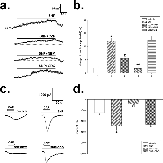
Figure 7.

The effect of SNP on membrane potential and capsaicin-induced inward current (Icap) in cultured NG neurons. (a) Representative membrane potential traces of the NG neurons after administration of SNP (0.16 mM), or co-application of SNP and NEM (5 mM) or ODQ (10 µM). The filled bars on the trace represent the administration of corresponding drugs. (b) Summary values show that application of SNP (0.16 mM) depolarized the membrane potential of NG neurons gradually. CZP (50 µM) or NEM (5 mM) blocked the effect of SNP, while ODQ (10 µM) had no effect. n = 7 in each column, *P < 0.05 vs vehicle, # P < 0.05 vs SNP group, ## P < 0.01 vs SNP group, Dunn’s post hoc test after a one-way ANOVA. (c) Representative traces of Icap after incubation of SNP (0.16 mM), or co-application of SNP and NEM (5 mM) or ODQ (10 µM). NEM eliminated the activation of Icap caused by SNP. Open bars on the trace represent the application of capsaicin for 60 s, and the filled bars indicate the pretreatment of corresponding drugs. (d) Summary values show that administration of SNP (0.16 mM) increased Icap. This effect was abolished by NEM, but not ODQ. n = 7 in each column, *P < 0.05 vs vehicle, ## P < 0.01 vs SNP group, Dunn’s post hoc test after a one-way ANOVA.
