
Figure 3.
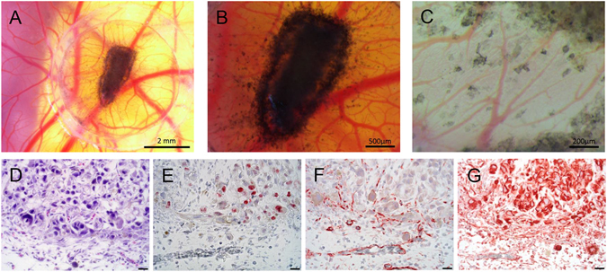
Ex ovo CAM assay: MUG-Mel2 onplants formed highly pigmented tumors within the silicone ring on the CAM surface after three days of incubation. Avian vessels developed radially towards the onplants (A: 10x magnification, B: 25x magnification), loco-regional metastases with solitary evading tumor cells surrounded by newly formed blood vessels were present (C: 63x magnification). Morphological analysis of hematoxylin/eosin stained sections revealed strong interaction of MUG-Mel2 cells with the CAM mesenchyme and invasion of tumor cells from the primary onplant site into the surrounding CAM tissue (D). MUG-Mel2 cells are mitotic active (Ki-67 staining) (E), tumor-driven neoangiogenesis was detected with anti-desmin (F). MUG-Mel2 cells express the tumor biomarker vimentin (G) (400x magnification, scale bar = 20 µm).