
Figure 5.
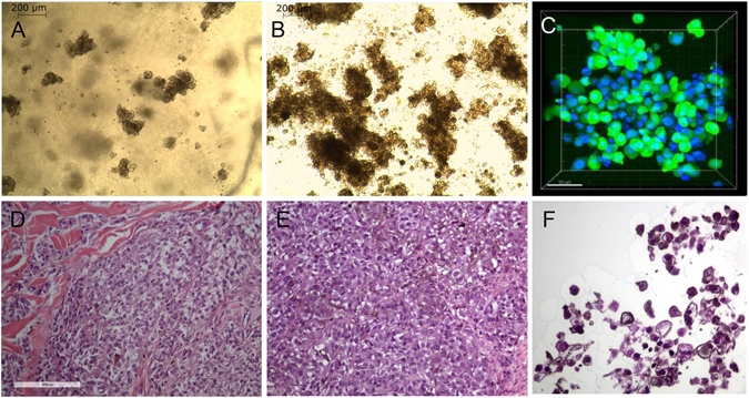
Morphology of MUG-Mel2 in 3D cultures. Cells formed spheroids in nanofibrillar cellulose (NFC) after two days in a concentration of 0.4% (A: 50x magnification), after five days in a concentration of 0.4% (B: 50x magnification); intensive brown staining was observed. Calcein staining (green) revealed the viability of the spheroids, nuclei were counterstained with DAPI (blue) (C). HE staining of human tumor tissue (D), tumor xenotransplantat (E), and 3D culture of MUG-Mel2 cells showed typical melanoma morphology and pigmented cytoplasm (F).