
Figure 7.
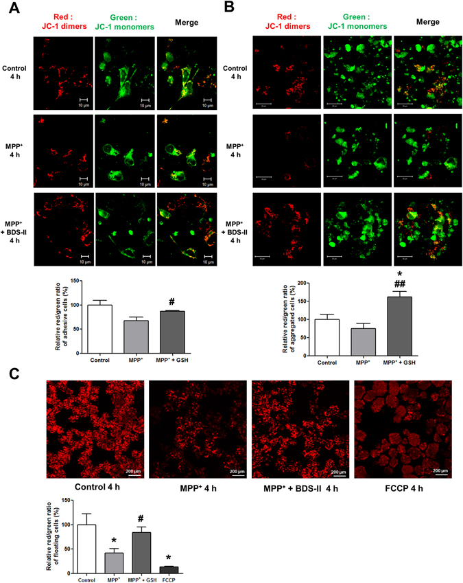
BDS-II blocked MPP+-induced MMP depolarization in SH-SY5Y cells (A) JC-1 staining revealed that BDS-II prevented the depolarization of mitochondrial membrane potential induced by 1 mM MPP+ in SH-SY5Y cells. Red JC-1 dimers represent normal mitochondrial membrane potential. Green JC-1 monomers represent depolarized mitochondrial membrane potential. Red dimers and green monomers were co-localized in control cells. When cells were treated with 1 mM MPP+, most of the cells showed only green monomers. When we pretreated with 100 nM BDS-II for 10 min followed by 4 h of 1 mM MPP+ and 100 nM BDS-II treatment, red dimers and green monomers co-localized again, but many cells also exclusively exhibited red dimers. (B) SH-SY5Y cells can form aggregates of undifferentiated cells. BDS-II also prevented the depolarization of mitochondrial membrane potential induced by 1 mM MPP+ in aggregated SH-SY5Y cells. Red dimers and green monomers were detected in the control and MPP+ + BDS-II groups, whereas almost all of the red dimers disappeared in the MPP+ group. (C) SH-SY5Y cells also grew into floating clumps of cells. Red JC-1 dimers indicated that 100 nM BDS-II rescued SH-SY5Y cells from MMP depolarization induced by 1 mM MPP+ treatment. FCCP (100 µM) was used as a positive control. All of the experiments were performed in triplicate or quadruplicate, and representative images are shown.