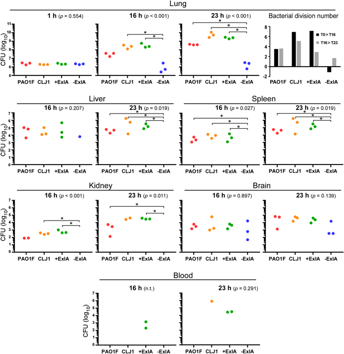
Figure 1.

Pulmonary infection: bacterial load in the lung and dissemination in various mouse organs. Mouse lungs were infected with 2.5 × 10⁶ bacterial suspensions of PAO1F, CLJ1, PAO1FΔT3SS::exlBA (+ExlA), PAO1FΔT3SS::empty vector (−ExlA). Organs (lung, spleen, liver, kidney, brain and blood) were isolated at 1, 16 and 23 h.p.i., as indicated, were homogenized and serial dilutions of the homogenates were plated onto agar plates to determine the CFU per organ. Of note, no bacteria were found outside of the lung at 1 h.p.i. Data are represented by solid circles on logarithmic scales. Three mice were used per strain and per time point. Statistical differences were calculated using Kruskal-Wallis’s test and probabilities are indicated between parenthesis in each condition; n.t., not testable; pairwise comparisons were established with Holm-Sidak’s post-hoc test: *p < 0.05. In the lungs, the bacterial division rates between 1 and 16 h.p.i. (T1 > T16), and between 16 and 23 h.p.i. (T16 > T23), were calculated for the different strains and plotted (upper right).
