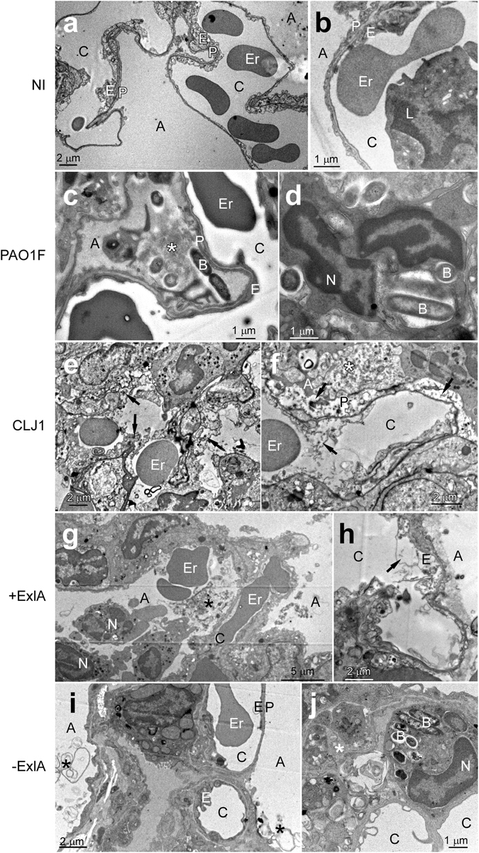
Figure 2.

Electron micrographs of infected lungs. (a–j) Mice (n = 2 per strain) were infected with 5 × 10⁶ bacteria from PAO1F, CLJ1, PAO1FΔT3SS::exlBA (+ExlA) or PAO1FΔT3SS::empty vector (−ExlA) strains, or were uninfected (NI), as indicated (2 images per condition). Mice were euthanized at 18 h.p.i. and lungs were isolated and prepared for electron microscopy. Abbreviations: A, alveolus; B, bacterium; C, capillary; E, endothelial cell; Er, erythrocyte; L, leukocyte; N, neutrophil; P, pneumocyte, *intra-alveolus material: mucus, cellular debris, surfactant. Arrows indicate endothelial or epithelial necrosis.
