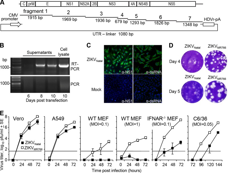
FIG 1 .
De novo generation of infectious ZIKVNatal by CPER. (A) Schematic representation of CPER assembly with seven synthetic DNA fragments covering the entire genome of ZIKVNatal with ~22-nt overlapping ends. The fragments were mixed with the UTR linker, which contained the CMV promoter followed by the first 22 nt of ZIKVNatal and, at the other end, the last 22 nt of ZIKVNatal, an HDVr site, and a poly(A) tail (pA). After CPER, the products were transfected into Vero cells. (B) On the posttransfection days indicated, RNA was isolated from Vero cell supernatants and a cell lysate and subjected to RT-PCR with fragment 5-specific primers (RT-PCR). To demonstrate that the bands were not due to contaminating DNA, a control (RT minus) PCR was performed in parallel (PCR). (C) Vero cells were infected with ZIKVNatal (culture supernatant from day 10 posttransfection), and mock-infected cells were used as controls. At 3 days postinfection, Vero cells were analyzed by immunofluorescent-antibody staining with anti-NS1 (4G4) and anti-dsRNA (3G1) antibodies (green) and DAPI counterstain (blue). (D) Plaque morphology following infection of Vero cells with ZIKVNatal and ZIKVMR766. Vero cells were fixed and stained with crystal violet on the postinfection days indicated. (E) Growth kinetics of ZIKVNatal and ZIKVMR766 in the cell lines indicated. Infection was performed at an MOI of 0.1, unless otherwise indicated. Virus titers in the culture supernatants were determined by plaque assays on Vero cells. The data and standard errors (SE) shown are from six independent experiments with Vero cells, A549 cells, and WT MEFs (MOI of 1) and three independent experiments with WT MEFs (MOI of 0.1), IFNAR−/− MEFs, and C6/36 cells. The horizontal line represents the limit of detection (50 PFU/ml).

