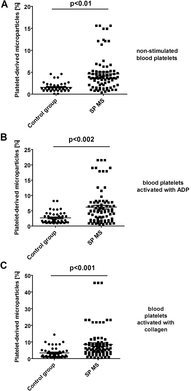
Fig. 4.
Formation of platelet-derived microparticles in control (a) and agonist-stimulated platelets: ADP 20 µM (b) and collagen 20 µg/ml (c) in whole blood samples obtained from SP MS patients and healthy controls. The fraction of platelet-derived microparticles was distinguished from platelets (CD61-positive objects) based on their size and granularity on the forward light scatter (FSC) vs. side light scatter (SSC) plots. CD61-positive objects with FSC lower than 102.3 were characterized as PMPs. In each sample, 10 000 CD61-positive objects (platelets) were measured. Statistical analysis was performed using Mann–Whitney U test

