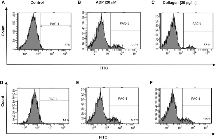
Fig. 6.
Representative histograms of the expression level of active form of GPIIb/IIIa on non-stimulated platelets (shown as control) (a, d) and on stimulated platelets with ADP (b, e) or collagen (c, f) in whole blood samples obtained from SP MS patients (d–f) and healthy controls (a–c). The blood platelets in whole blood were distinguished based on the expression of CD61 conjugated with PerCP. For each sample, 10 000 CD61-positive objects (platelets) were acquired. The blood platelets were labeled with monoclonal antibody PAC-1 (conjugated with FITC) against the active form of GPIIb/IIIa

