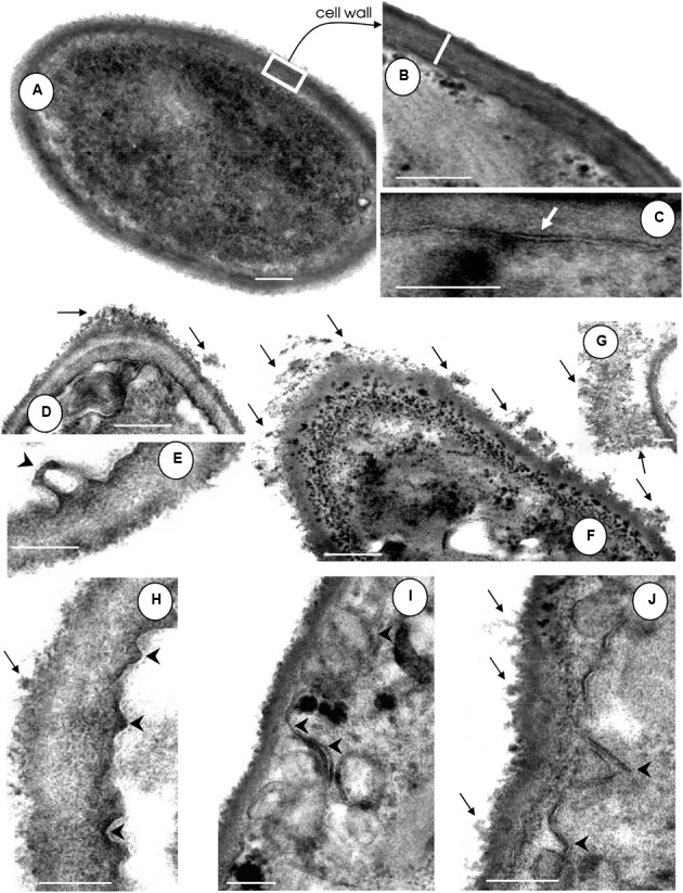
FIGURE 2.

Effect of HIV-PIs on the ultrastructure of F. pedrosoi conidial cells. (A–C) Untreated and (D–J) HIV-PI-treated (100 μM for 24 h) conidial cells were processed and analyzed by transmission electron microscopy. Control conidial cells present a dense cytoplasm (A) with a distinct and compact cell wall (B, white bar shows the wall thickness) and well-delineated plasma membrane (C, white arrow). Black arrows show the releasing of electron dense and amorphous material from the cell surface and black arrowheads indicate the invaginations of the plasma membrane with consequent withdrawal from the cell wall following treatment of conidia with indinavir (D,E), nelfinavir (F,G), ritonavir (H) and saquinavir (I,J). Scale bars: (A,D,F), 0.4 μm; (B–J), 0.25 μm.
