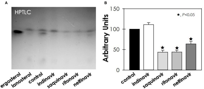
FIGURE 4.
Effect of HIV-PIs on the sterol synthesis of F. pedrosoi conidial cells. (A) Representative image of untreated (control) and HIV-PI-treated (100 μM for 24 h) conidial cells. For sterol analysis, the lipid extracts were loaded into HPTLC silica plates, separated using a mixture of solvents (hexane-ether-acetic, 80:40:2) and revealed by spraying a solution containing ferric chloride, water, acetic acid and sulfuric acid, which revealed the sterol as red-violet color spots. (B) Densitometric analyses of the lanosterol bands, expressed in arbitrary units. Symbols (★72) indicate the experimental systems considered statistically significant from the control (P < 0.05, Student’s t-test).

