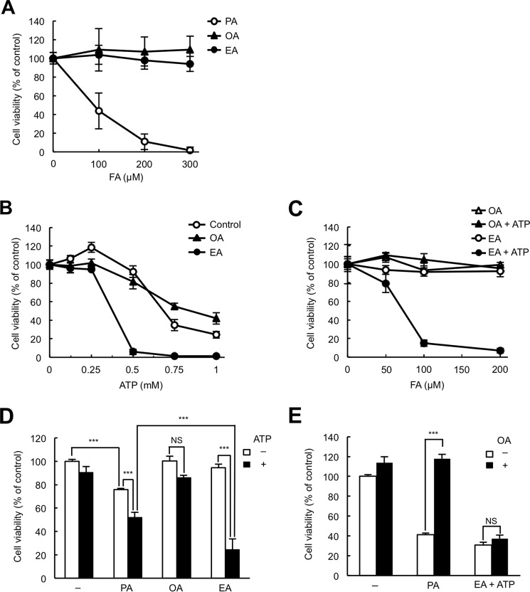
Figure 1.
EA promotes extracellular ATP-induced cell death. A, RAW264.7 cells were treated with the indicated concentrations of PA, OA, or EA for 24 h and subjected to a cell viability assay. The data shown are the mean ± S.D. FA, fatty acid. B, RAW264.7 cells were pretreated with or without 200 μm fatty acid as indicated for 12 h, stimulated with various concentrations of ATP for 6 h, and then subjected to a cell viability assay. The data shown are the mean ± S.D. C, RAW264.7 cells were pretreated with various concentrations of OA or EA for 12 h, stimulated with 0.5 mm ATP for 6 h, and then subjected to a cell viability assay. The data shown are the mean ± S.D. D, RAW264.7 cells were pretreated with 100 μm PA, OA, or EA for 12 h, stimulated with 0.5 mm ATP for 6 h, and then subjected to a cell viability assay. The data shown are the mean ± S.D. Significant differences were determined by one-way ANOVA followed by Tukey-Kramer test. ***, p < 0.001; NS, not significant. E, RAW264.7 cells were pretreated with 200 μm PA or EA in the presence or absence of 100 μm OA for 12 h, and then EA-pretreated cells were stimulated with 0.5 mm ATP for 6 h and subjected to a cell viability assay. The data shown are the mean ± S.D. Significant differences were determined by one-way ANOVA followed by Tukey-Kramer test. ***, p < 0.001.

