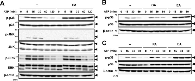
Figure 2.
EA enhances p38 activation induced by extracellular ATP. A and B, RAW264.7 cells were pretreated with 200 μm OA or EA for 12 h and then stimulated with 0.5 mm ATP for the indicated periods. Cell lysates were subjected to immunoblotting with the indicated antibodies. Arrowheads indicate bands corresponding to the indicated proteins, and the asterisk indicates a nonspecific band. C, RAW264.7 cells were treated with 100 μm PA or EA for 12 h, and stimulation and immunoblot analysis were performed as in A and B. Arrowheads indicate bands corresponding to the indicated proteins.

