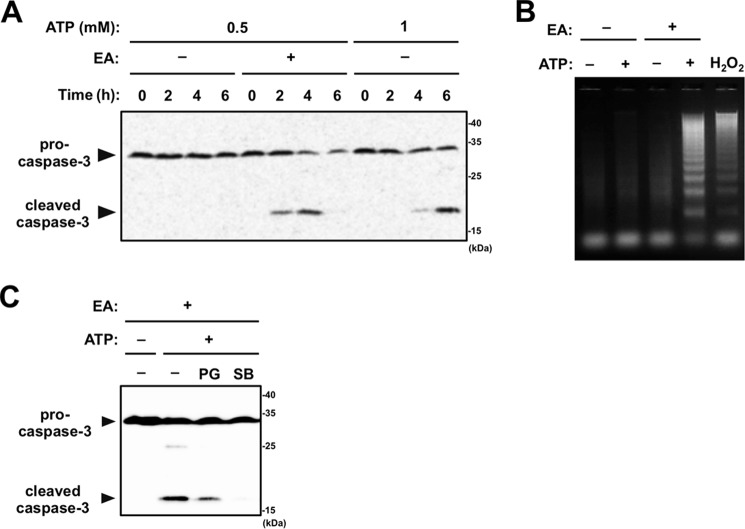
Figure 4.
EA promotes apoptosis induced by extracellular ATP. A, RAW264.7 cells were pretreated with or without 200 μm EA for 12 h and then stimulated with 0.5 or 1 mm ATP for the indicated periods. Cell lysates were subjected to immunoblotting with an anti-caspase-3 antibody. B, RAW264.7 cells were pretreated with or without 200 μm EA, stimulated with vehicle, 0.5 mm ATP, or 0.5 mm H2O2 for 4 h, and then subjected to a DNA fragmentation assay. C, RAW264.7 cells were pretreated with 200 μm EA for 12 h in the presence of either the antioxidant PG or the p38 inhibitor SB203580 (SB), stimulated with 0.5 mm ATP for 4 h, and then subjected to immunoblotting with an anti-caspase-3 antibody.

