Figure 5.
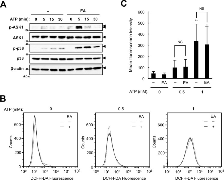
EA enhances ATP-induced ASK1 activation without increased ROS generation. A, RAW264.7 cells were pretreated with or without 200 μm EA for 12 h and then stimulated with 0.5 or 1 mm ATP for the indicated periods. Cell lysates were subject to immunoblotting with the indicated antibodies. Arrowheads indicate bands corresponding to the indicated proteins. B and C, RAW264.7 cells were pretreated with or without 200 μm EA for 12 h, treated with 20 μm DCFH-DA for 30 min, and then stimulated with 0.5 mm ATP for 5 min. The amount of intracellular ROS was analyzed by FACS analysis, and a representative FACS plot (B) and mean ± S.D. (C) of four independent experiments are shown. Significant differences were determined by one-way ANOVA followed by Tukey-Kramer test. *, p < 0.05; **, p < 0.01 (versus control without ATP stimulation); NS, not significant.

