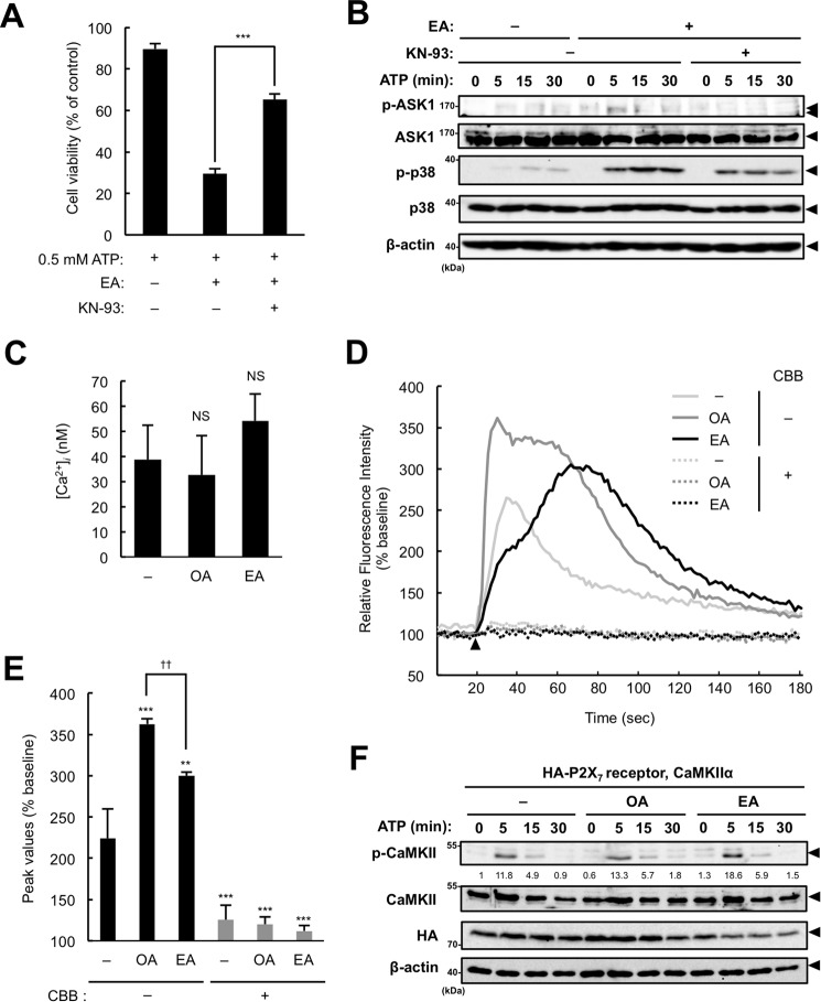
Figure 6.
CaMKII participates in the EA-mediated enhancement of ASK1 activation. A, RAW264.7 cells were pretreated with 200 μm EA for 12 h, treated with the CaMKII inhibitor KN-93 (10 μm) 30 min before stimulation with 0.5 mm ATP for 6 h, and then subjected to a cell viability assay. Data are presented as the percentage of viability of the cells treated with 200 μm EA without ATP stimulation and shown as mean ± S.D. Significant differences were determined by one-way ANOVA followed by Tukey-Kramer test. ***, p < 0.001. B, RAW264.7 cells were pretreated with 200 μm EA for 12 h and then treated with the CaMKII inhibitor KN-93 (5 μm) 30 min before stimulation with 0.5 mm ATP for the indicated periods. Cell lysates were subjected to immunoblotting with the indicated antibodies. Arrowheads indicate bands corresponding to the indicated proteins. C–E, RAW264.7 cells were pretreated with 200 μm EA for 12 h, loaded with Fluo-4/AM for 1 h, and then subjected to measurements of basal intracellular free Ca2+ concentrations ([Ca2+]i, mean ± S.D.) (C) and ATP-induced Ca2+ influx (D and E). The cells were stimulated with 0.5 mm ATP at 18 s after starting measurements (arrowhead), and the fluorescence was monitored intermittently for 3 min. Representative data of three independent experiments are shown as the percentage of basal fluorescent intensity (D), and the average peak values of the relative fluorescence intensity are shown as mean ± S.D. Significant differences were determined by one-way ANOVA followed by Tukey-Kramer test. NS, not significant (versus control) (C); **, p < 0.01; ***, p < 0.001 (versus control without CBB); ††, p < 0.01 (E). F, HEK293A cells were transfected with the HA-P2X7 receptor and CaMKII, pretreated with 200 μm OA or EA for 12 h, and then stimulated with 3 mm ATP for the indicated periods. Cell lysates were subjected to immunoblotting with the indicated antibodies. The values below the p-CaMKII blot indicate the relative phosphorylation levels of p-CaMKII normalized with total CaMKII levels. Arrowheads indicate bands corresponding to the indicated proteins.

