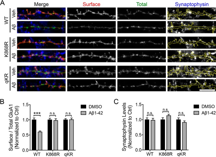
Figure 3.
GluA1 ubiquitination is necessary for the Aβ-induced reduction of surface AMPARs. A, cortical neurons were transfected with pH-GluA1 constructs, either WT or ubiquitin-deficient mutants as indicated, at DIV12. qKR contains quadruple mutations of Lys-813, Lys-819, Lys-822, and Lys-868 in the GluA1 C-terminal tail into arginines. At DIV14, neurons were treated with 5 μm Aβ or DMSO (Veh) for 1 h and incubated with anti-GFP antibodies prior to fixation to visualize surface pH-GluA1 expression (red). Total pH-GluA1 expression was determined with the endogenous GFP signal (green), whereas the level of synaptophysin was visualized by immunostaining with anti-synaptophysin antibodies (blue). Scale bar, 10 μm. The effects of Aβ on the levels of surface receptor expression (B) and synaptophysin (C) were quantified as surface/total receptor ratios and as total synaptophysin staining per dendritic area, respectively, and normalized to DMSO controls (Ctrl). Data represent the mean of two independent experiments (Mann-Whitney test; ***, p < 0.001; n.s., not significant; n = 30). Error bars represent S.E.

