Figure 6.
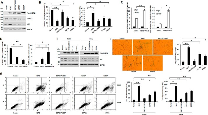
The phosphorylation at Ser-372/Ser-380 by Pim-1 is indispensable for HBP1-induced premature senescence and apoptosis. A, phosphorylation at Ser-372/Ser-380 is required for the regulation of DNMT1 and p16 by HBP1. The protein levels of HBP1, DNMT1, and p16 were measured by Western blotting in 2BS cells transfected with vector, HBP1, S372A/S380A, S372A, and S380A individually. B, phosphorylation at Ser-372/Ser-380 is required for the transactivation of DNMT1 and p16 by HBP1. Relative activities of HBP1 and associated mutants on the native DNMT1 or p16 promoter were determined by luciferase reporter gene assay. HEK293T cells were cotransfected with 0.1 μg of the indicated reporters and 0.8 μg of HBP1 or mutant expression plasmids. Error bars represent S.D. *, p < 0.05; **, p < 0.01. C, Pim-1 enhances the binding of HBP1 to DNMT1 or p16 promoter. HEK293T cells were transfected with HBP1 or HBP1 + Pim-1. A ChIP assay was used to test the binding of exogenous HBP1 to the endogenous DNMT1 or p16 promoter. Error bars represent S.D. *, p < 0.05; **, p < 0.01. D, Pim-1 enhances the transactivation of HBP1 on DNMT1 or p16 promoter. HEK293T cells were cotransfected with the indicated reporters and HBP1 or HBP1 + Pim-1 expression plasmids. Error bars represent S.D. *, p < 0.05; **, p < 0.01. E, the phosphorylation at Ser-372/Ser-380 is required for the induction of Bax by HBP1. U2OS and HeLa cells were transfected with vector, HBP1, S372A/S380A, S372A, or S380A individually. After 48-h transfection, Bax protein expression was analyzed by Western blotting. F, phosphorylation at Ser-372/Ser-380 contributes to HBP1-induced SA-β-gal staining. 2BS cells (PD20) were stably transfected with vector, HBP1, S372A/S380A, S372A, or S380A through lentiviral infection, respectively, and then cells were stained for SA-β-gal. Error bars represent S.D. *, p < 0.05; **, p < 0.01. G, phosphorylation at Ser-372/Ser-380 contributes to HBP1-induced apoptosis. U2OS and HeLa cells were transfected with vector, HBP1, S372A/S380A, S372A, or S380A individually. After 48-h transfection, cell apoptosis rates were measured by FACS. Error bars represent S.D. *, p < 0.05; **, p < 0.01.

