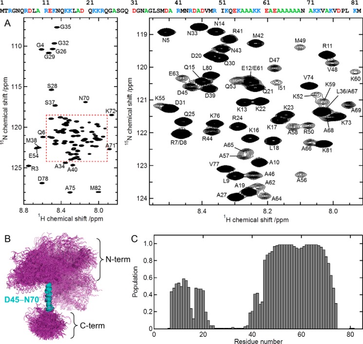
Figure 2.
MOAG-4 forms a malleable structure with α-helical propensity. A, 1H-15N HSQC spectrum of MOAG-4 (left), and a zoomed region (right). The amino acid sequence of MOAG-4 is shown on the top, using the following color scheme: basic (blue), acidic (red), alanine (green). B, ensemble of MOAG-4 structures. The structures are superimposed on the α-helical residues Asp45-Asn70. C, the population of α-helical structure is calculated from the MOAG-4 ensemble.

