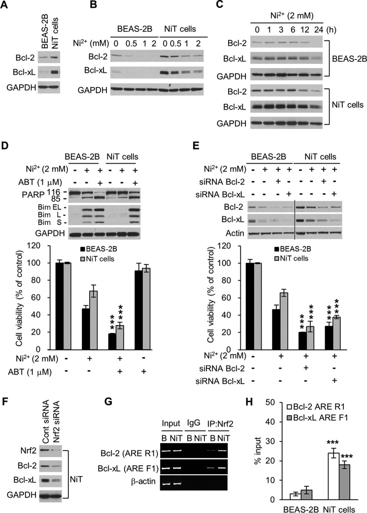
Figure 5.
Nrf2 mediates the high expression of antiapoptotic proteins Bcl-2 and Bcl-xL in NiT cells. The expression levels of Bcl-2 and Bcl-xL in BEAS-2B and NiT cells were evaluated by Western blot analysis (A). BEAS-2B and NiT cells were exposed to various concentrations of Ni2+ (0–2 mm) for 24 h (B) or to Ni2+ (2 mm) for various lengths of time (0–24 h) (C), and the levels of Bcl-2 and Bcl-xL were assessed by Western blot analysis. To demonstrate inhibition, BEAS-2B and NiT cells were incubated with Ni2+ (2 mm) for 24 h in the presence or absence of ABT-263 (1 μm); the expression levels of Bcl-2 and Bcl-xL were assessed by Western blot analysis, and cell viability was assessed using MTT (D). BEAS-2B and NiT cells were transfected with siRNA to knock down Bcl-2 and Bcl-xL; the expression levels of Bcl-2 and Bcl-xL were assessed by Western blot analysis, and cell viability was assessed using MTT (E). NiT cells were transfected with siRNA to knock down Nrf2, and the expression levels of Bcl-2 and Bcl-xL were assessed by Western blot analysis (F). The binding of Nrf2 to the Bcl-2 and Bcl-xL promoters was examined by ChIP analysis. The Bcl-2 ARE R1 or Bcl-xL ARE F1 regions were analyzed by conducting normal real-time PCR (G) or quantitative real-time PCR (H) assays with primers specific for the ARE-containing region of the promoters. Data are presented using the percent input method and are normalized to each control. ***, p < 0.001 indicates a significant difference from exposure to Ni2+ only or to vehicle control, as determined by ANOVA and Scheffe's test. GAPDH and β-actin were used as loading controls. Bim EL, extra long; Bim S, short; Bim L, long.

